
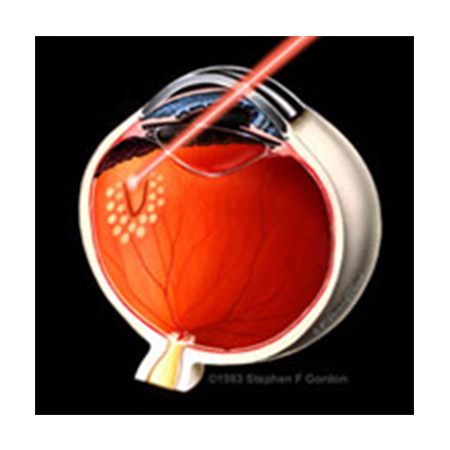
A laser is a powerful beam of light which, combined with ophthalmic equipment and lenses, can be focused on the retina. All lasers cause a certain amount of controlled damage in order to elicit the desired effect. Small bursts of the laser can be used to seal leaky blood vessels, destroy abnormal blood vessels, seal retinal tears, and destroy abnormal tissue in the back of the eye.
Laser treatment is performed in the office, and usually requires no anesthesia other than an eyedrop. The procedure may take a few minutes, or can last up to half an hour, depending on the type of treatment needed. Most patients do not require a patch or medications following retinal laser, and can resume normal activities immediately.
Several different laser procedures are used in the treatment of the following retinal diseases:
Patients with disorders affecting the blood vessels of the macula, in particular diabetic retinopathy and retinal vein occlusion, often require this procedure. The laser decreases the leakage from damaged blood vessels, helping to preserve normal retinal thickness and function. The procedure is painless and does not take long to perform.
When abnormal retinal blood vessel growth (neovascularization) occurs in diseases such as proliferative diabetic retinopathy or retinal vein occlusion, laser must be applied in a "scatter" pattern to large areas of the peripheral retina. These areas of the retina have poor blood flow (ischemia), and are responsible for releasing growth factors that cause the neovascularization. Untreated, retinal neovascularization often leads to bleeding in the eye (vitreous hemorrhage), traction retinal detachment, and/or neovascular glaucoma. After laser is applied, the blood vessels tend to stabilize or regress. Since this treatment affects the function of the retinal periphery, some patients will recognize decreased peripheral and night vision. The size of the pupil and the central vision may also be affected in some patients.
Abnormal blood vessel membranes can grow beneath the retina in age-related macular degeneration and other similar conditions. There are three main types of laser treatment which are used to eliminate these membranes:
When a retinal tear or a small retinal detachment occurs, laser treatment may be applied to prevent further accumulation of fluid beneath the retina and minimize the risk of a vision-threatening retinal detachment. The laser is applied around the retinal defect. Over the course of a few weeks, the treated area develops a scar, which forms a tight seal between the retina and the underlying tissue. This procedure is sometimes performed around weak areas in the retina, such as lattice degeneration, in patients who may be at higher risk for retinal detachment. Laser retinopexy is often performed in conjunction with surgical treatments for retinal detachment, such as vitrectomy or pneumatic retinopexy.
Copyright ©2020 Aditya EYE Hospital || All rights reserved. Design by Sevy Inc